Difference between revisions of "Bacteria"
savitanaik (talk | contribs) |
savitanaik (talk | contribs) |
||
| Line 35: | Line 35: | ||
==Reference Books== | ==Reference Books== | ||
| − | #[http://ncert.nic.in/NCERTS/textbook/textbook.htm?hesc1=2-18 NCERT Text Book | + | #[http://ncert.nic.in/NCERTS/textbook/textbook.htm?hesc1=2-18 NCERT Text Book Chapter on Micro organism -Friend and foe] |
2. PUC Ist year and 2nd Year Text Books. | 2. PUC Ist year and 2nd Year Text Books. | ||
Revision as of 17:58, 30 August 2013
| Philosophy of Science |
While creating a resource page, please click here for a resource creation checklist
Concept Map
Error: Mind Map file Bacteria.mm not found
Textbook
To add textbook links, please follow these instructions to: (Click to create the subpage)
Additional information
Useful websites
3.1 Useful websites.:
Reference Books
2. PUC Ist year and 2nd Year Text Books.
Teaching Outlines
Concept Bacteria
1. Habitat and size of Bacteria.
2. Classification of bacteria (based on shapes)
3. Structure of bacteria.
Brief Notes:
Habitat and size of bacteria:
Bacteria (/bækˈtɪəriə/ ( listen); singular: bacterium) are a large domain of prokaryotic microorganisms.
Bacteria are found everywhwere. - even on you !
Approximately 2,000 species have been identified, many of them living in conditions that would destroy other organisms.
 They have been found in the almost airless reaches of the upper atmosphere,
10 km (6 mi) below the surface of the ocean, in frozen soil, and on rocks in hot springs.
 Some bacteria produce a resting stage, the endospore, which is the most resistant living thing
known and can be killed only by boiling in steam under pressure for many hours.
Size of bacteria:
Thirty trillion bacteria of average size weigh about 28 g (1 oz). Bacteria are measured in microns (0.001 micrometer, about 0.00004 in) and most types range from 0.1 to 4.0 microns in width and 0.2 to 50 microns in length.
2. Classification of bacteria on the basis of shapes:
Content: Teaching Outlines.
Bacteria are neither plant nor animal. Both bacteria and plants have rigid cell walls, but unlike plants,
most kinds of bacteria move about and use organic foods for energy and growth; only a few use photosynthesis.
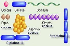
On the basis of their shapes,bacteria may be grouped into three main type;
*the rod-shaped bacilli, which often have small whiplike structures known as Flagella that propel the organism;
*the spherical cocci (singular coccus), which may grow in chains (streptococci, or strep germs,"
as in strep throat) or which may clump together like   a bunch of grapes Istrphylococci);
*and the comma or spiral shape spirilla and prirochetes (one of which is the cause of syphilis).
other kind of bacteria, the mycoplasmas, have no rigid cell walls and consequently are called pleuropneumonialike organisms,
because they cause a contagious pneumonia in cows and human beings.
3) Structure of Bacteria :
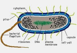
Structure of Bacterial Cell
Bacterial is very small, simple unicellular organisms with a length varying from 2 to 5.
On the basis of shape bacteria are either coccus (spherical), bacillus (rod shape), spirillum (spiral) or vibro (comma) types.
On the basis of arrangement of flagella, these may be atrichous (flagellum absent)
monotrichous (one flagellum at one end), leptotrichous (bunch of flagella on one side), amphitrichous (flagella on whole surface).
Internal structure:
Cell wall:
Bacterial cell is surrounded by a prominent cell wall constituted by polysaccharides, lipids and proteins.
The cell wall is permeable to water and ions of small molecules.
Slime layer and capsule:
Some bacterial cells are completely enveloped by a slimy layer, which is relatively thick to form the capsule. Capsulated bacteria are more harmful. Capsule protects the cell from antibodies and desiccation.
Mesosomes:
Mesosomes take part in aerobic respiration and it is found in gram positive bacteria. The protoplasm is either transparent or granular.
Protoplasm:
Below the cell wall, the plasma membrane is present. Plasma membrane at certain points forms coiled invaginations called Mesosomes.
Cytoplasm:
Cytoplasm is composed of complex proteins, lipids and mineral, nucleic acid and water.
Glycogen is the reserve food material. It contains 70S type of ribosomes.
Other organelles like endoplasmic reticulum, mitochondria; Golgi complex, etc. are absent.
However, in photosynthetic bacteria some pigments are present (bacterio chlorophyll).
Nucleoid:
Bacteria are prokaryotes, there is no well-organised nucleus. Nuclear membrane and nucleolus are absent.
At the center there is a clear zone called nucleoid where only a single naked chromosome (without histone protein, only DNA)
is present in a very much coiled form.
Episomes
In addition to the DNA in some bacterial cells an additional circular DNA is present in the cytoplasm. It is called as episome or plasmid.
Learning objectives
Students will--
- Understand the characteristics of monera .
- Describe the habitat of bacteria .
- Classify bacteria on the basis of shapes.
- Identify the structure and morphology of bacteria.
Notes for teachers
Teacher can introduce the lesson on bacteria by giving a brief introduction about monera and prokaryotes.
 ( Internal link the lesson Click here File:1. introduction to monera.pdfIntroduction to monera for reference).
4. Teacher Should be familiar with the meaning of following terms related to bacteria.
Words to Know
1.Aerobic bacteria: Bacteria that need oxygen in order to live and grow.
2.Anaerobic bacteria: Bacteria that do not require oxygen in order to live and grow.
3. Bacillus: A type of bacterium with a rodlike shape.
4. Capsule: A thick, jelly-like material that surrounds the surface of some bacteria cells.
5. Coccus: A type of bacterium with a spherical (round) shape.
6. Decomposers: Bacteria that break down dead organic matter.
7. Fimbriae: Short, hairlike projections that may form on the outer surface of a bacterial cell.
8. Fission: A form of reproduction in which a single cell divides to form two new cells.
9. Flagella: Whiplike projections on the surface of bacterial cells that make movement possible.
10. Pasteurization: A process by which bacteria in food are killed by heating the food to a particular temperature for some given period of time.
11. Pili: Projections that join pairs of bacteria together, making possible the transfer of genetic material between them.
12. Prokaryote: A cell that has no distinct nucleus.
13. Spirilla: A type of bacterium with a spiral shape.
14. Spirochetes: A type of bacterium with a spiral shape.
15. Toxin: A poisonous chemical.
16. Vibrio: A type of bacterium with a comma-like shape. 0
Teacher can develop the lesson by --
- first explaining bacteria is a micro organism and is a prokaryote.
- showing different images of bacteria ( Refer Link about images of habitat of bacteria in google search) showing the bacteria is present everywhere.
- Demonstrating the below experiment of milk changing into curds and by showing the figure about bacterial infection on man.
Thus students can get clear idea that bacteria are found everywhere.

Activity No 1
- Materials/ Resources needed
Milk , lemon
- Prerequisites/Instructions,
This experiment can be conducted to Level : Std 6th ,7th. 8th &9th
Time Required: 20 Minutes.
- Multimedia resources
Charts/ Diagrams
- Website interactives/ links/ simulations
Wesite interactions/links/ simulations
 Image credit-- Resources needed are Refer Link about images of habitat of bacteria in google search  to show that bacteria is present everywhere. [1]
Ppt. on structure of Bacteria File:Bacterial Structures (1).pdf
- Process/ Developmental Questions
Procedure:Add lemon juice to a cup of milk . See the changes. Milk changes into curds.
How did this change take place? (Lactococcus lactis is a microbe classified informally as a Lactic Acid Bacterium
because it ferments milk sugar (lactose) to lactic acid.)
- Evaluation
Evaluation: What changes do you find when lemon juice is added to milk? Is there change in the taste? Why did this change take place?
- Question Corner
Activities to students for evaluation (CCE)
1. Browse the web and prepare the presentation on habitat and types of bacteria.
2. Draw the different shapes of bacteria on drawing sheet , name them and display in class room.
3. Why does the milk get spoilt during summer? Does it get spoilt if we boil milk? If not, why?
4. Similarly which food items get spoilt soon? Why?
Questions corner.After going through the images and seeing the picture, what can we conclude about habitat of bacteria?
Are they found even on your body?!
Project Ideas
Do this activity at home: About a cup of milk is taken in a vessel & kept for 1-2days. Similarly A lemon is cut into halves and kept for 2days.
After 2 days students will observe the changes and give the reasons for the change.)
Fun corner
Usage
Create a new page and type {{subst:Science-Content}} to use this template


